Videos & More
Sample Videos from Netter's Online Dissection Modules by UNC at Chapel Hill
-
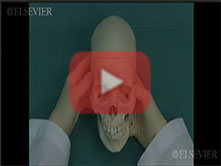
Video 26. Osteology of the Head and Neck: Step 4. Introduction to the skull -
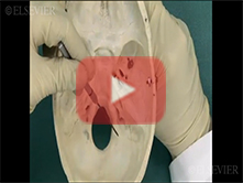
Video 27. Osteology of the Head and Neck: Step 23. Foramina of the posterior cranial fossa -
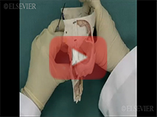
Video 28. Osteology of the Head and Neck: Step 25. Paranasal sinuses -
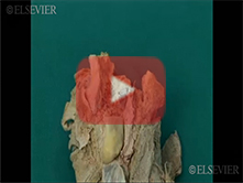
Video 29. Oral Cavity and Infratemporal Fossa: Step 6. Temporomandibular joint and attachment of the lateral -
Video 30. Oral Cavity and Infratemporal Fossa: Step 12. Submandibular ganglion: innervation of the sublingua -
Video 31. Vertebral Column and Its Contents: Step 1, Approach to a laminectomy -
Video 32. Vertebral Column and Its Contents: Step 6, Sacral canal and sacral hiatus; dorsal rami of sacral s -
Video 33. Vertebral Column and Its Contents: Step 3, Spinal cord, conus medullaris, internal filum terminale, -
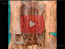
Video 34. Vertebral Column and Its Contents: Step 2, Epidural space and dura mater -
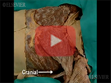
Video 35. Anterior Body Wall, Mediastinum and Heart : Step 1, Anterior thoracic wall and intercostal nerves -
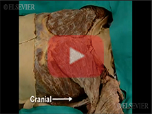
Video 36. Anterior Body Wall, Mediastinum and Heart : Step 3, External and internal intercostal muscles -
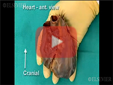
Video 37. Coronary Circulation and Internal Structure of the Heart: Step 1, Surface of the heart, right coron -
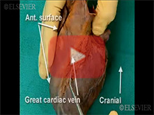
Video 38. Coronary Circulation and Internal Structure of the Heart: Step 3, Great cardiac vein and coronary -
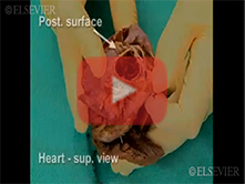
Video 39. Coronary Circulation and Internal Structure of the Heart: Step 9, Aortic valve, aortic sinuses and -
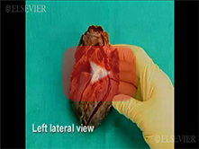
Video 40. Coronary Circulation and Internal Structure of the Heart: Step 10, Opening the outflow tract -
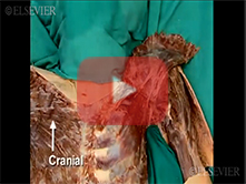
Video 41. Anterior Shoulder, Pectoral Region, Breast and Brachial Plexus: Step 4, Subclavian vein and subclav -
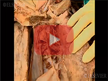
Video 42. Lungs and Pleural Cavity: Step 4, Left vagus nerve, phrenic nerve and ligamentum arteriosum -
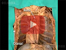
Video 43. Posterior Mediastinum, Azygos System: Step 2, Relationship of the esophagus to the tracheal bifurca -
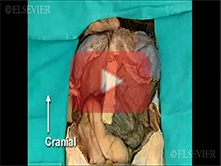
Video 44. Anterior Abdominal Wall and Abdominal Viscera in situ: Step 12, Abdominal viscera in situ -
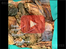
Video 45. Celiac Trunk, Superior Mesenteric Vessels and Related Viscera: Step 6, Primary and secondary branch -
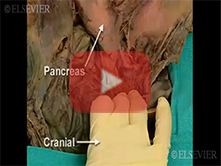
Video 46. Celiac Trunk, Superior Mesenteric Vessels and Related Viscera: Step 8, Branches of the splenic and -
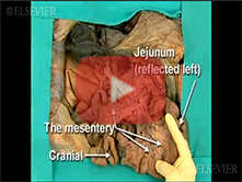
Video 47. Celiac Trunk, Superior Mesenteric Vessels and Related Viscera: Step 12, Jejunum and ileum -
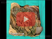
Video 48. Diaphragm, Kidneys and Posterior Abdominal Wall: Step 4, Ureter and gonadal veins -
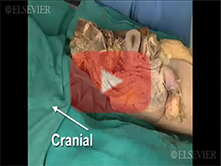
Video 49. Pelvic Vasculature and Autonomic Innervation: Step 2, Pelvic splanchnic nerves -
Video 50. Posterior Mediastinum, Azygos System: Step 7, Confluence of the azygos vein and the superior vena c -
Video 51. Female Pelvic Viscera: Step 1, Pelvic viscera in situ -
Video 52. Female Pelvic Viscera: Step 6, Uterosacral and cardinal ligaments -
Video 53. Female Pelvic Viscera: Step 4, Uterus -
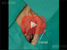
Video 54. Male External Genitalia and Perineum: Step 12, Root and bulb of the penis -
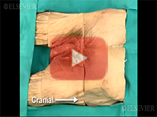
Video 55. Male External Genitalia and Perineum: Step 1, Removal of skin from the perineum -
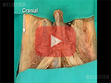
Video 56. Male External Genitalia and Perineum: Step 11, Structure of the penis: erectile bodies, vasculature -
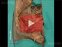
Video 57. Male Pelvic Viscera: Step 9, Superficial perineal space and perineal membrane -
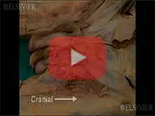
Video 58. Male External Genitalia and Perineum: Step 5, External and internal spermatic fascia; cremaster mus -
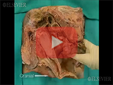
Video 59. Male Pelvic Viscera: Step 6, Anatomical features of the rectum -
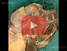
Video 60. Pelvic Vasculature and Autonomic Innervation: Step 3, Sacral splanchnic nerves -
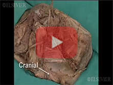
Video 61. Pelvic Vasculature and Autonomic Innervation: Step 10, Branches of the internal iliac artery -
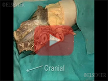
Video 62. Pelvic Vasculature and Autonomic Innervation: Step 4, Middle sacral artery, common iliac artery and -
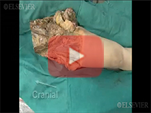
Video 63. Somatic Nerves of the Greater and Lesser Pelvis; Pelvic Diaphragm: Step 1, Ilioinguinal and iliohyp -
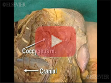
Video 64. Somatic Nerves of the Greater and Lesser Pelvis; Pelvic Diaphragm: Step 9, Pudendal nerve and its r -
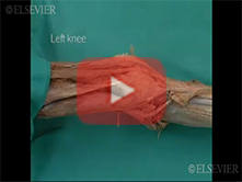
Video 65. Knee: Step 4, Medial (Tibial) collateral ligament -
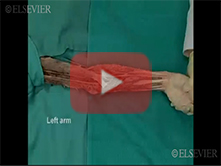
Video 66. Flexor Surface of the Forearm: Step 2, Median nerve and brachial artery in the cubital fossa -
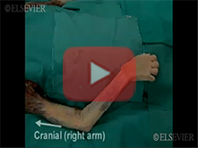
Video 67. Flexor and Extensor Compartment of the Arm and Shoulder Joint: Step 1, Olecranon bursa, brachial fa -
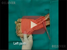
Video 68. Palm : Step 1, Approach to the palm, palmaris longus, palmar aponeurosis -
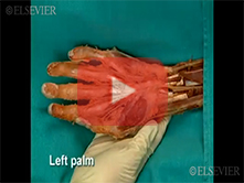
Video 69. Palm: Step 6, Common flexor synovial sheath, ulnar bursa -
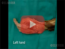
Video 70. Extensor Surface of the Forearm and Dorsum of the Hand: Step 7, Extensor apparatus; lumbrical and i -
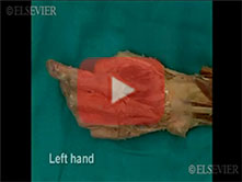
Video 71. Extensor Surface of the Forearm and Dorsum of the Hand: Step 8, Nail bed, distal interphalangeal (D -
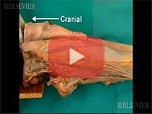
Video 72. Medial and Anterior Thigh: Step 4, Sartorius muscle; medial and lateral intermuscular septa -
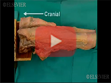
Video 73. Medial and Anterior Thigh: Step 3, Iliotibial band and tensor fascia lata -
Video 74. Knee: Step 3, Lateral (Fibular) collateral ligament -
Video 75. Knee: Step 5, Patella, patellar ligament and patellar retinaculum -
Video 76. Anterior and Lateral Compartments of the Leg, Dorsum of the Foot; Ankle Joint: Step 7, Contents of -
Video 77. Plantar Surface of the Foot: Step 1, Plantar aponeurosis -
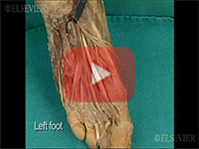
Video 78. Plantar Surface of the Foot: Step 4, Third layer -
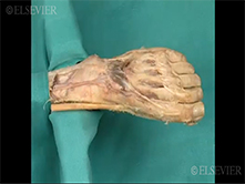
Video 79. Anterior and Lateral Compartments of the Leg, Dorsum of the Foot; Ankle Joint: Step 10, Dorsal inte
