Videos & More
Sample Videos from Netter's Online Dissection Modules by UNC at Chapel Hill
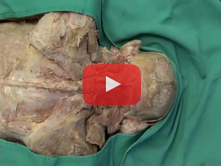
UNC 1. Posterior Neck and Suboccipital Region: Step 5. Suboccipital triangle 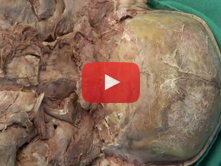
UNC 2. Posterior Neck and Suboccipital Region: Step 6. Exposure of the posterior arch of C1 and related str 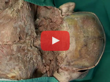
UNC 3. Posterior Neck and Suboccipital Region: Step 7. Vertebral artery and suboccipital nerve 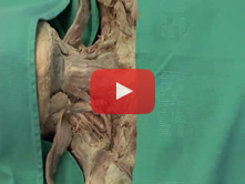
UNC 4. Anterior Neck: Step 6. Infrahyoid and digastric muscles; submandibular gland 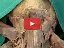
UNC 5. Anterior Neck: Step 7. Subdivisions of the anterior triangle 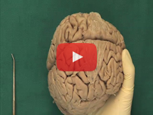
UNC 6. Brain: Step 1. Cerebral hemispheres, lobes, fissures, gyri and sulci 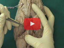
UNC 7. Brain: Step 2. Diencephalon: mammillary bodies, pituitary gland, tuber cinereum, optic nerves, and p 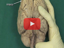
UNC 8. Brain: Step 3. Mesencephalon: cerebral peduncles, interpeduncular fossa, oculomotor nerve, trochlear 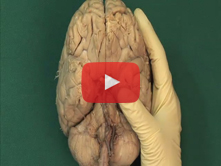
UNC 9. Brain: Step 4. Metencephalon: pons and cerebellum and associated cranial nerves 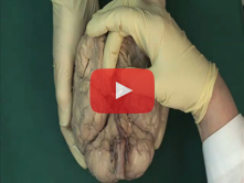
UNC 10. Brain: Step 5. Medulla oblongata: olive, pyraminds, apertures, and choroid plexus of the fourth vent 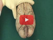
UNC 11. Brain: Step 6. Cerebral arterial circle (of Willis) 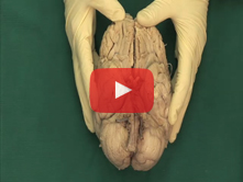
UNC 12. Brain: Step 7. Middle, anterior and posterior cerebral arteries 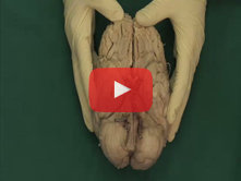
UNC 13. Brain: Step 8. Midsagittal section of the brain 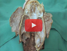
UNC 14. Brain: Step 9. CN III, V, and VI in the floor of the cranial cavity; trigeminal cave and ganglion; m 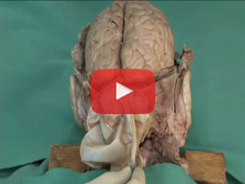
UNC 15. Cranial Cavity and Dural Venous Sinuses: Step 10. Reflection of the tentorium cerebelli and removal 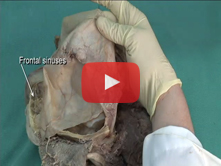
UNC 16. Cranial Cavity and Dural Venous Sinuses: Step 11. Dural venous sinuses 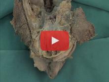
UNC 17. Eye and Orbit I: Step 2. Frontal nerve 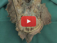
UNC 18. Eye and Orbit I: Step 3. Superior oblique muscle; trochlear and lacrimal nerves 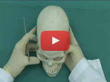
UNC 19. Osteology of the Head and Neck: Step 7. Cranial sutures 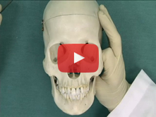
UNC 20. Osteology of the Head and Neck: Step 19. Foramina in the base of the skull 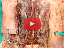
UNC 21. Vertebral Column and Its Contents: Step 2. Epidural space and dura mater 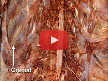
UNC 22. Vertebral Column and Its Contents: Step 3. Spinal cord, conus medullaris, internal filum terminale, 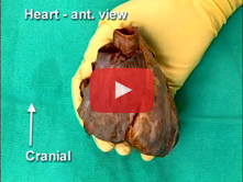
UNC 23. Coronary Circulation and Internal Structure of the Heart: Step 4. Internal anatomy of the right atri 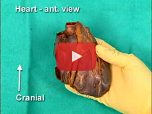
UNC 24. Coronary Circulation and Internal Structure of the Heart: Step 5. Internal anatomy of the right vent 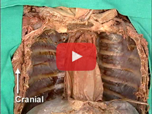
UNC 25. Posterior Mediastinum, Azygos System: Step 1. Vagus and recurrent laryngeal nerves 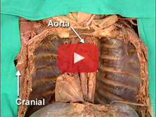
UNC 26. Posterior Mediastinum, Azygos System: Step 2. Relationship of the esophagus to the tracheal bifurcat 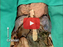
UNC 27. Anterior Abdominal Wall and Abdominal Viscera in situ: Step 11. Ligaments of the liver 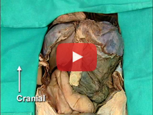
UNC 28. Anterior Abdominal Wall and Abdominal Viscera in situ: Step 12. Abdominal viscera in situ 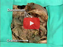
UNC 29. Stomach, Duodenum, Portal System, and Inferior Mesenteric Artery: Step 8. Internal anatomy of the du 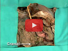
UNC 30. Stomach, Duodenum, Portal System, and Inferior Mesenteric Artery: Step 9. Duct system within the pan 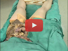
UNC 31. Female Pelvic Viscera: Step 1. Pelvic viscera in situ 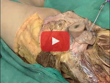
UNC 32. Female Pelvic Viscera: Step 5. Pelvic ligaments and uterine tube 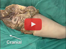
UNC 33. Somatic Nerves of the Greater and Lesser Pelvis; Pelvic Diaphragm: Step 6. Lumbosacral trunk and sac 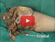
UNC 34. Somatic Nerves of the Greater and Lesser Pelvis; Pelvic Diaphragm: Step 7. Obturator internus fascia 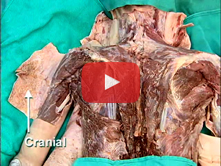
UNC 35. Posterior Shoulder: Step 2. Rhomboid major, rhomboid minor, and levator scapulae muscles; accessory 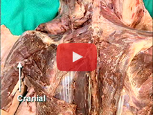
UNC 36. Posterior Shoulder: Step 3. Innervation of the rhomboid and latissimus dorsi muscles; the dorsal sca 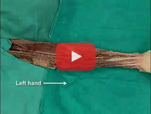
UNC 37. Flexor Surface of the Forearm: Step 4. Pronator teres and muscular branches of the median nerve 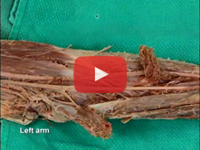
UNC 38. Flexor Surface of the Forearm: Step 5. Sectioning of first layer of flexor muscles 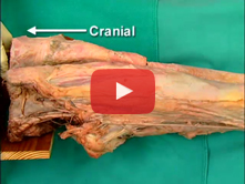
UNC 39. Medial and Anterior Thigh: Step 4. Sartorius muscle; medial and lateral intermuscular septa 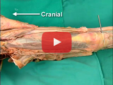
UNC 40. Medial and Anterior Thigh: Step 5. Quadriceps femoris muscle 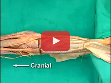
UNC 41. Popliteal Fossa, Knee Joint, and Posterior Compartment of the Leg: Step 5. Triceps surae and its inn 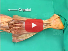
UNC 42. Popliteal Fossa, Knee Joint, and Posterior Compartment of the Leg: Step 6. Contents of the deep post
